This same technical principle is what inspires
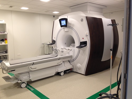
Magnetic Resonance, the most modern procedure to date, which based on given properities of magnetic fields enables users not only to obtain remarkably high-quality anatomical images but also to perform spectroscopic analyses of the various tissues of the human body.
All of the above has been complmented by the ongoing surge in therapeutic procedures, which, under the heading of Interventional Radiology, range from the performance of a cystogram to the drainage of an abscess, the dilation of an artery, or the embolization of an aneurysm.

HIFU
In HM HOSPITALS we have the latest thecnology
The historical and technical evolution of the specialty, and the continuous incorporation of new technology obtained in some cases with the help of physical agents outside the class of ionizing radiation, has allowed the concept of Imaging Services to be conceived for new Radiology departments. These
Imaging Services have paved the way for the practice of integrated radiology, or in other words combined diagnostic radiology, which is simply the application of the method to support the technique, and which includes selecting the most suitable procedure (radiological technique), recongizing and analysing the signs (semiology), and interpreting these correctly.
Thus, the method will be an intelligent sequence of actions for achieving the objectives of the radiological examination. The decade of the 1980s, which saw the complete development of most modern techniques, brought with it and interesting dilemma for the specialty: the choice between the convenience of organizing Radiology according to the model of organs and systems, in line with the traditional divison of medicine and surgery; and doing so based on the use of the various existing techniques.
While the effectiveness of both options is debatable depending on where the activity is carried out, most developed countries generally tend towards the planning and organization of
Diagnostic Radiology by organ and system, as this arrangement is of greater benefit to the different medical- surgical speicalties.
Leaving aside the above, which in practice still has operational drawbacks in many countries, for years it has been recognized that there are three traditional sphereres of knowledge,
Pediatric Radiology, Neuroradiology and Vascular and Interventional Radiology, which have been operating within Diagnostic Radiology Department seperately and in their own right.

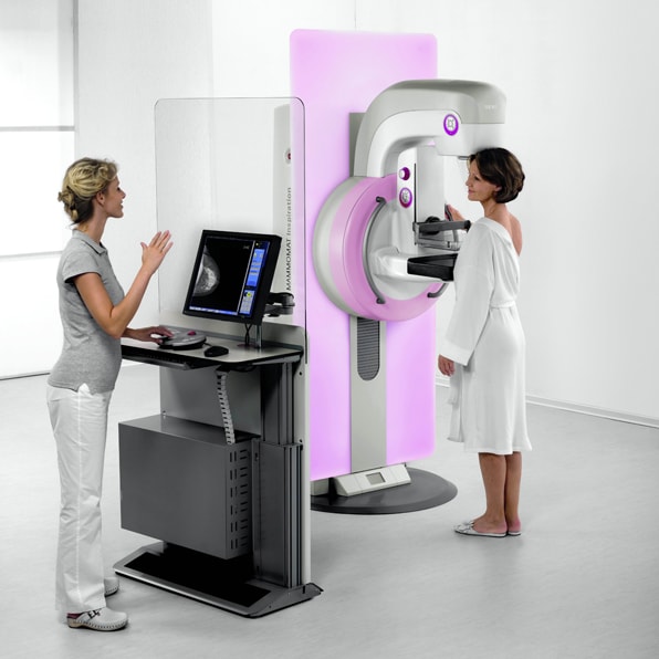
LAST GENERATION OF MAMMOGRAPHER

AQUILION ONE
HM SANCHINARRO

FIRST MRI VERTICAL SCANNER
Radiology Service
In addition to conventional and remote-controlled radiology devices, the Diagnostic Imaging Service has:
• 160 multislice CT scan (Toshiba Aquilion) with 80 detectors. It can perform coronary artery examinations and the most complex studies in a few minutes.
• Closed high field MRI (RNM MAGNETOM ESPREE Siemens 1.5 T) - High magnetic field equipment with a compact magnet that is wider and shorter than conventional resonances. Indicated for claustrophobic and obese patients, it provides high quality images.
• Breast Pathology Unit. In this unit, we offer a digital mammogram with tomosynthesis (SELENIA DIMENSIONS). We have also other systems for advanced imaging such as sterotaxy, and the latest generation breast ultrasound (ESAOTE MyLabEight XP), an ultrasound system that defines a new standard in image quality for a better diagnosis.
• EOS – a low radiation 3D vertical radiology: Vertical radiological diagnostic system that allows obtaining 2D images and 3D reconstructions, having the patient standing up or even sitting in a transparent chair to X-rays.
• Prostate Fusion Biopsy: Currently, it is the most effective technique for the diagnosis of prostate cancer thanks to the combination of images obtained by conventional transrectal ultrasound and those of magnetic resonance, a prostate mapping is generated. This information is very precious for the specialist when the biopsy is made. In this way, samples are collected only from areas detected as tumorous or suspicious.
• PHILIPS ECOGRAPH (AFFINITY 70 DS): Conventional ultrasound (muscle, transference, soft tissue…)
• PORTABLE ECOCARDIO (PHILIPS COMPACT EXTREM CX-50 CARD)
• GYNECOLOGICAL ECOGRAPHY (GENERAL ELECTRIC-VOLUSON 730 Pro): Abdominal/vaginal gynecological ultrasound and 3D ultrasound.
• ORTHOPANTOMOGRAPH (ORTHORALIX 9200 GENDEZX): For dental X-rays and skull X-rays.
• FIBROSCAN (FIBROSCAN PROBE M +) - Study of the elasticity of the liver (fibrosis fracture) replacing liver biopsy.
Appointments or enquiries
Contact us
We will assist you at any hospital in Spain.